
Tirzepatide and Semaglutide Structures: What Cryo-EM Can Show
Cryo-electron microscopy can show how a peptide occupies a receptor complex and which molecular contacts are supported by the reconstructed density. A structure of tirzepatide or semaglutide does not, by itself, measure binding duration, signaling strength or biological performance. Those questions require additional experiments, and an unresolved region should not be mistaken for a chemically absent region.
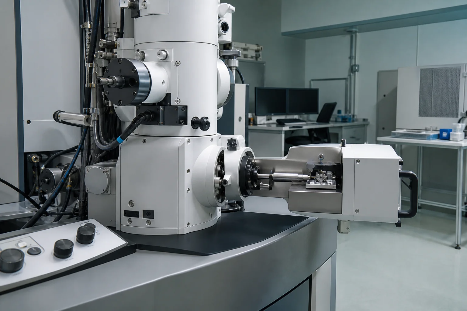
The distinction matters because a molecular image looks unusually conclusive. A colored ribbon appears complete, a dashed contact looks permanent, and two overlapping structures can look equivalent. Reading the underlying evidence reveals a more useful question: which parts of this explanation were observed, which were modeled, and which were tested independently?
Start with the complex, not just the peptide name
Single-particle cryo-EM reconstructs a three-dimensional density map from many images of particles. Researchers then fit and refine a molecular model against that map. The map and the fitted coordinates are related outputs, but they are not the same object: the coordinates express an interpretation of the experimental density.
A peptide-bound receptor structure also contains an experimental context. A receptor associated with a particular signaling partner is a selected complex, rather than an inventory of every state that receptor visits in a cell. Our introduction to GLP-1 receptor models explains the target terminology; here, the important extra detail is the actual assembly used for structural work.
For example, the deposited tirzepatide–GLP-1R structure, PDB 7RGP, identifies an electron-microscopy experiment involving the human GLP-1 receptor and additional components. The deposition provides access to the structure, experimental information and validation material. It is a place to inspect what was modeled, not a certificate for a separately supplied peptide sample.
When reading a figure, expand its short label into a sentence: this is this ligand, bound to this receptor construct, in this prepared complex. That sentence prevents a GLP-1R image from silently becoming evidence about GIPR, or a G-protein-associated assembly from becoming evidence about every other receptor partner.
A real tirzepatide example: missing density is informative
In Sun and colleagues' 2022 structural study, tirzepatide was examined with GIPR and GLP-1R. The authors resolved peptide residues 1–32, while residues 33–39 and the linker–fatty acid moiety were unresolved. They supplemented the structures with molecular dynamics and studies of modified peptide analogs.
That distinction is more informative than a picture drawn with every atom present. An unresolved attachment has not been shown to be absent; the structural experiment has not located it reliably in the reported reconstruction. Conversely, drawing the missing attachment into a model does not make its position an experimental observation.
The practical reading habit is to preserve three separate labels: resolved in the reconstruction, added for modeling, and tested in a follow-up experiment. A reader can then understand why a structural hypothesis is plausible without assuming that every element of it was directly visualized.
This matters particularly for Tirzepatide, where a peptide name alone does not tell a reader which portion of the molecule a displayed structural model includes. Compare the molecular description with the figure legend before treating an apparently short ribbon as a complete chemical representation.
Semaglutide shows why one consensus structure is not the whole story
Zhang and colleagues' 2021 semaglutide and taspoglutide study combined GLP-1R–Gs structures with three-dimensional variability analysis. The peptides had broadly similar interactions to GLP-1, while the analysis revealed differences in movements of the peptides and receptor. The semaglutide linker region also required more careful interpretation than a single consensus map could provide.
This creates a useful distinction between structural similarity and behavioral identity. Similar overall binding arrangements can coexist with differences in the conformations represented in the data. A reader should therefore ask whether a displayed model summarizes one class of particles, a consensus reconstruction or an analysis of variability.
The Semaglutide name identifies the molecule being discussed. It does not mean that all semaglutide figures, including older fragment or isolated-domain structures, depict the same molecular assembly. Check the construct and modification state before comparing pictures from different papers.
Four claims that need different evidence
| Claim being considered | Evidence to inspect | Limit to retain |
|---|---|---|
| The peptide occupies this region of the receptor | Density map and fitted model | The claim concerns the reconstructed complex |
| A proposed contact matters for function | Structural observation plus a suitable perturbation and response measurement | A changed response may have more than one explanation |
| A region moves between conformations | Variability analysis or other dynamic evidence | A static ribbon alone does not establish the movement |
| One peptide produces a larger biological response | Matched functional measurements | Structural resemblance or contact count is insufficient |
This table is an editorial reading aid, not a new structural analysis. It keeps the type of evidence next to the question that evidence can answer. For the measurements in the final row, see our separate guide to receptor assay readouts.
How to read a contact claim without overinterpreting it
Suppose a figure highlights a peptide residue near a receptor side chain. First ask whether the local density supports the proposed orientation. Then ask whether the authors changed that residue and measured a relevant consequence. Finally, check whether the experiment helps distinguish a local interaction from a broader change in folding, expression or molecular behavior.
These questions do not make structural work less valuable. They show where the argument becomes strongest. A convincing explanation connects observations across methods, rather than expecting one image to establish every part of a mechanism.
Molecular dynamics adds another layer. A simulation can explore a proposed system over time, but its starting structure and assumptions remain part of the result. In a summary, use “the simulation suggested” for a simulated behavior and “the experiment measured” for an observed response. Avoid blending both into “the structure proved.”
Within the GLP-1 peptide collection, this approach helps readers keep related materials distinct. The category groups a research area; it does not imply that one receptor structure explains all included molecules.
Limits and a useful stopping point
This article examines selected structural papers, not every published receptor state. Purification, complex stabilization, reconstruction and model building all affect the evidence available for interpretation. A structural contact cannot establish an outcome in another biological system, and these publications did not analyze a NEXTWAVE PEPTIDES product lot.
A strong final summary can be short: name the complex, identify the supported observation, explain the follow-up evidence, and state what remains unresolved. When comparing the two molecules more broadly, the tirzepatide and semaglutide research overview provides the surrounding context. Keep the structural conclusion as specific as the experiment that supports it.