
LL-37 Antimicrobial Assays: Why Salt, Medium, and Endpoint Change the Reading
An LL-37 antimicrobial result belongs to its test organism, strain, medium, ionic conditions, incubation time, peptide preparation, and endpoint. A value from one assay is not a fixed property that can be transferred across every microbial species or laboratory environment. The correct reading keeps the assay conditions in the same sentence as the result.
What is LL-37 in these studies?
LL-37 is a 37-residue human cathelicidin-derived peptide studied in laboratory models of host defense and microbial response. Even when two papers use the name LL-37, a comparison still needs the reported sequence, preparation, concentration basis, storage, and assay design. Full-length LL-37, a fragment, and an analogue are different research materials.
Related research tool: For a transparent assay plan, keep concentration arithmetic in the Laboratory Concentration and Dilution Calculator and replicate allocation in the Research Sample Planner.
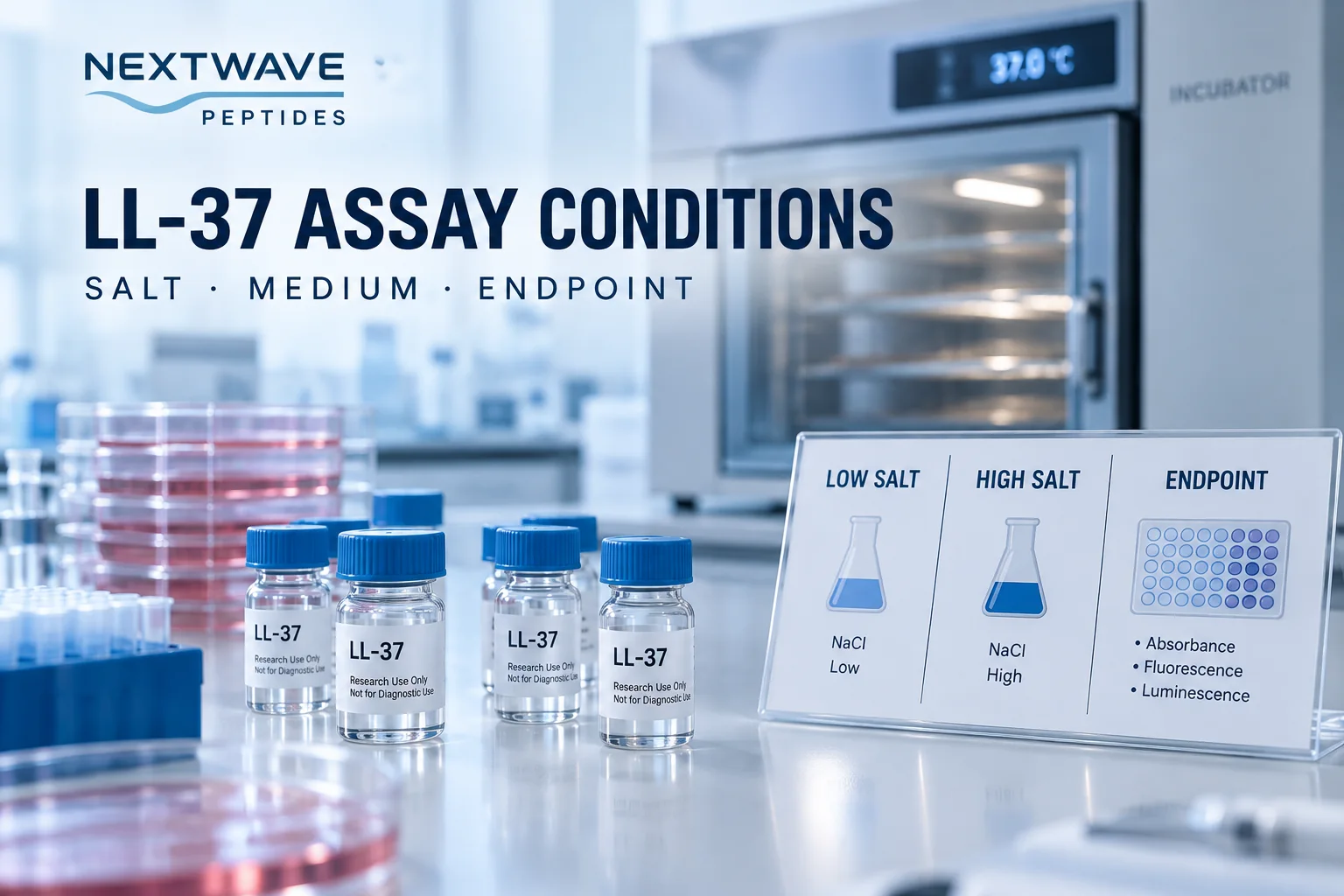
The name of the peptide is only one experimental variable. Ionic strength can change electrostatic interactions, medium components can bind or shield a peptide, and different organisms have different surfaces and susceptibility patterns. The endpoint then determines what “activity” means in that experiment.
One paper can show several condition-dependent readings
Turner and colleagues compared LL-37 with other antimicrobial peptides using radial diffusion and a broth microdilution approach. The study reported broad activity across several organisms, while also finding that some organism-medium combinations behaved differently in the presence of 100 mM sodium chloride. Methicillin-resistant *Staphylococcus aureus*, *Proteus mirabilis*, and *Candida albicans* were reported as resistant under that salt condition but susceptible in low-salt media. Other tested organisms did not follow the same pattern. Turner et al. (1998)
That result does not support the shortcut “salt turns LL-37 off.” It supports a narrower statement: in this study, salt sensitivity depended on the organism and experimental conditions. The same paper also used membrane-permeabilization and lipopolysaccharide-binding measurements, which answer different questions from growth inhibition.
What each endpoint actually measures
| Endpoint | What is observed | Why it should not be treated as a synonym |
|---|---|---|
| Radial diffusion | A zone produced as peptide diffuses through a defined gel environment | Diffusion and gel composition contribute to the readout |
| Broth MIC | The lowest tested concentration meeting the study's growth-inhibition definition | It does not directly count surviving organisms |
| Viable count | Colony-forming organisms after exposure and plating | Timing and recovery conditions affect the result |
| Luminescence or fluorescence | A reporter signal used as a proxy for microbial state | The proxy requires validation against the biological outcome |
| Membrane permeabilization | Movement of a reporter across a membrane | Permeabilization is a mechanistic readout, not automatically cell death |
When two papers report “potency” but use different rows from this table, their numbers should not be ranked without qualification.
Ionic strength is not a single universal switch
Travis and colleagues tested mammalian cathelicidin-derived peptides at ionic strengths of 25 and 175 mM against several bacteria. For LL-37 and the other peptides, responses varied by organism, peptide, and assay. The study used both a luminescence readout and viable counts for *Pseudomonas aeruginosa*, and compared those data with radial-diffusion measurements. This design is useful because it shows that ionic conditions and endpoint selection travel together in the result. Travis et al. (2000)
The experiment also compared LL-37 with cathelicidins and derived peptides from other species. Results for CAP18, SMAP29, or a truncated peptide cannot be reassigned to full-length LL-37 merely because the molecules share a family or structural theme. The material identity must stay attached to every row of a comparison.
A defensible LL-37 comparison record
For each experiment, capture these fields before comparing values:
- peptide sequence, terminal form, source, and preparation;
- organism, strain, growth phase, and starting inoculum;
- complete medium or buffer description;
- sodium concentration, total ionic strength, divalent ions, and serum where reported;
- peptide concentration basis and exposure time;
- temperature and mixing conditions;
- endpoint definition and detection method;
- positive, negative, vehicle, and medium controls;
- replicate count and how variability was reported;
- whether the result is inhibition, killing, permeabilization, or another outcome.
The same table should leave missing fields blank or label them “not reported.” Filling gaps with values from another experiment creates a comparison that looks complete but no longer describes either source accurately.
Why medium composition can change interpretation
Microbiological media are active parts of an assay. Protein, salts, nutrients, pH, and other components can change peptide availability, microbial growth, reporter behavior, and the physical environment at the cell surface. A low-salt buffer designed to reveal peptide activity and a nutrient-rich standardized broth may produce different values without implying that the molecular identity changed.
This is why “physiological conditions” is too vague for an evidence table. A paper should provide the actual formulation or enough detail to reproduce it. If the formulation is absent, the limitation should appear next to the result.
Connecting literature to material documentation
The KPV, LL-37, and Thymosin Alpha-1 guide keeps these peptide identities and literature objects separate. BPC-157 and TB-500 Evidence Boundaries provides a broader method for separating model, endpoint, and claim. The Research-Peptide Literature Matrix can store organism, medium, salt, time, and endpoint in the same row.
For wider catalog context, see Research Compounds. A catalog record can identify a supplied material and its documentation. It cannot inherit antimicrobial outcomes from a paper unless the material and assay relationship is demonstrated explicitly.
Research limitations
The cited work consists of in-vitro antimicrobial and mechanistic assays under specified laboratory conditions. These experiments do not establish clinical effects, dosing, safety, or performance of a catalog product. Results may change with organism, strain, medium, salt, serum, peptide preparation, incubation, and endpoint. The evidence supports condition-specific interpretation rather than a universal LL-37 activity value.
Frequently asked questions
Can MIC values from two LL-37 studies be compared directly?
Only after checking organism, strain, inoculum, medium, ionic conditions, incubation time, endpoint definition, and peptide identity. If those fields differ, a direct numerical ranking can mislead.
Does high salt always eliminate LL-37 activity?
No. Published results show organism- and condition-dependent behavior. A statement about salt must name the tested system and endpoint.
Are radial diffusion and broth MIC results equivalent?
No. Diffusion through a gel and growth inhibition in broth involve different physical environments and readout definitions. They can complement each other but should retain separate labels.
Does an LL-37 paper verify a product lot?
No. A paper describes its study material. A specific product lot requires its own identity and test documentation.